
Interventional Cardiologist
Dr. Manish Kumar Jha
MBBS, MD (General Medicine), DM (Cardiology)
Angiography, angioplasty and stents, heart attack and heart failure care.

An echocardiogram, or “2D echo”, uses ultrasound to show the heart’s chambers, valves and pumping in real time. It is one of the most common heart tests, uses no radiation and needs no special preparation. Echocardiography is done in the cardiology department at Charak Hospital, Hardoi Road, Lucknow.
Share your details — our coordinator will call you back to schedule a visit.
A small probe with gel is moved over the chest. It sends sound waves that bounce off the heart and form moving pictures on a screen. Colour Doppler shows the direction and speed of blood flow through the valves.
Measures ejection fraction (EF).
Checks for narrowing or leaking valves.
No radiation, safe in pregnancy.
Report usually the same day.
Common reasons include:
To check for heart failure or valve disease.
To see how the heart muscle is working.
To look at the valves.
To check heart function.
To check thickening of the heart.
To assess fitness for anaesthesia.
To check for heart defects.
After heart surgery or treatment.
Severe chest pain or breathlessness now? Do not wait for an outpatient echo. Come to the 24×7 emergency department or call 0522-666-4444. Call 0522-666-4444.
A standard echo needs no special preparation.
As usual, unless told otherwise.
Wear a two-piece outfit; you will be asked to uncover the chest. A gown is provided.
Previous echo, ECG and angiography reports.
If your doctor advises a different type of echo, the team will give you specific instructions.
Here is what to expect.
You lie on your left side. Small ECG stickers are placed on your chest. The cardiologist or technician moves the probe with gel over different areas of the chest and may ask you to breathe in, out or hold your breath.
Takes about 20 to 40 minutesYou may hear whooshing sounds of blood flowSlight pressure from the probeGeneral guidance — always follow the advice your own doctor gives you.
The percentage of blood pumped out with each beat. Around 55 to 70 percent is generally normal; your doctor will explain your value.
“Stenosis” means narrowing; “regurgitation” means leaking. Trace or mild leaks are common and often not significant.
Areas that move less may suggest damage from reduced blood supply.
MBBS, MD (General Medicine), DM (Cardiology)
Angiography, angioplasty and stents, heart attack and heart failure care.

MBBS, MD (General Medicine), DM (Cardiology)
Coronary angiography and angioplasty, chest pain, high BP and heart rhythm problems.
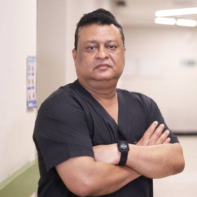
MBBS, MS, M.Ch (CTVS)
Bypass surgery (CABG), heart valve surgery and chest surgery.
A little preparation helps us help you faster.
Tick off as you pack:
Ask our team for the current echo charges. Outpatient tests may or may not be covered by your insurance policy.
*Coverage depends on your policy and the treatment. Our insurance desk can check this for you.
Call 0522-666-4444No. An ECG records the heart’s electrical activity; an echo is an ultrasound that shows the heart’s structure and pumping.
Not for a standard echo.
Yes. It uses sound waves, not radiation.
About 20 to 40 minutes.
Yes. It is commonly used to check for heart defects in children.
No matching term. Please ask your doctor at your next visit.
Bring your doctor’s prescription. Call 0522-666-4444.
Medical information disclaimer: This page provides general health information for patients and families. It is not a substitute for a consultation, diagnosis or treatment by a qualified doctor. Treatment decisions depend on each patient's individual condition. In an emergency, call 0522-666-4444 or go to the nearest emergency department.
About this page: Prepared by the Charak Hospital web team with the help of AI writing tools, using information from the Institute of Cardiac Sciences, Charak Hospital & Research Centre · Last updated: October 2026