
In This Article
What Is the ACL?
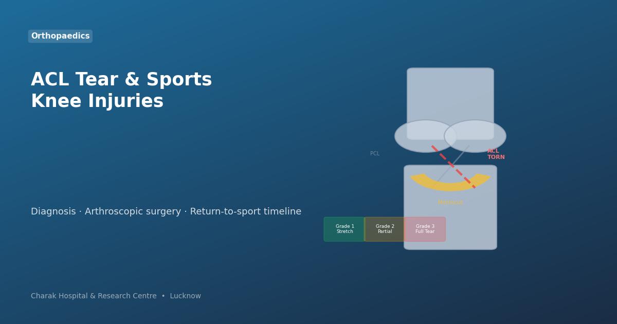
The knee has four main ligaments that hold it together — the ACL (Anterior Cruciate), PCL (Posterior Cruciate), MCL (Medial Collateral), and LCL (Lateral Collateral). The ACL runs diagonally through the centre of the knee, preventing the tibia (shinbone) from sliding forward on the femur (thighbone) and providing rotational stability.
When the ACL tears, the knee loses this stability — causing the characteristic "giving way" sensation, especially during cutting and twisting movements.
How ACL Tears Happen
About 70% of ACL injuries occur without contact — from a sudden deceleration, landing from a jump, or a sharp pivot. Common scenarios:
- Landing awkwardly from a jump (basketball, volleyball, kabaddi)
- Sudden change of direction at speed (football, hockey, badminton)
- Stopping abruptly from running
- Direct blow to the knee (contact sports)
- Hyperextension of the knee
At the moment of injury, many patients hear or feel a distinct "pop" in the knee, followed by rapid swelling within hours.
Symptoms of an ACL Tear
Audible or felt snap at the moment of injury.
Haemarthrosis (blood in joint) causes swelling within 2–6 hours.
Most athletes cannot continue playing immediately after an ACL tear.
Knee buckles unpredictably — especially on stairs, uneven ground, or twisting movements.
Pain along the joint line; difficulty bending or straightening the knee fully.
Joint swelling limits bending. Full range often returns as swelling subsides.
Grades of Ligament Injury
Diagnosis — Tests & Imaging
An experienced orthopaedic surgeon can diagnose an ACL tear clinically with a high degree of accuracy using:
- Lachman Test — the most sensitive clinical test for ACL tear. Examines forward displacement of the tibia.
- Anterior Drawer Test — classic ACL test performed with the knee at 90°.
- Pivot Shift Test — assesses rotational instability.
Investigations:
- MRI of the knee — gold standard for confirming ACL tear and detecting associated meniscus or cartilage injuries. Approximately 40–70% of ACL tears have concurrent meniscus tears.
- X-Ray — rules out associated fractures (tibial spine avulsion is common in younger patients).
Arthroscopic ACL Reconstruction
A torn ACL cannot heal on its own — it has no blood supply. For active patients (athletes, young people, those with physical jobs), arthroscopic ACL reconstruction is the standard of care.
The Procedure
Under anaesthesia, two small keyhole incisions are made. The arthroscope is inserted to visualise the joint. A graft — typically taken from the patient's own hamstring tendon (autograft) — is used to replace the torn ACL. The graft is fixed in bone tunnels using screws, positioned precisely to restore the original ACL anatomy. Total surgical time: approximately 60–90 minutes.
Graft Options
- Hamstring autograft — most common in India; taken from gracilis/semitendinosus. Minimal donor site morbidity.
- Patellar tendon autograft (BTB) — bone-to-bone healing, preferred for high-demand athletes. Slightly higher donor site pain.
- Allograft (cadaver tendon) — no donor site. Used when autograft is not feasible. Longer integration time.
Meniscus Injuries in Sport
The medial and lateral menisci are C-shaped cartilage discs that cushion and stabilise the knee. Meniscus tears are extremely common sports injuries — often occurring alongside ACL tears.
Symptoms: joint line pain (inner or outer knee), clicking or locking sensation, difficulty fully bending or straightening the knee, swelling.
Treatment: small tears in the avascular zone may heal conservatively. Larger tears, and tears in the vascular (outer) zone, are ideally repaired arthroscopically (meniscus repair) rather than removed — preserving the shock-absorbing function and reducing long-term arthritis risk. If meniscal tissue is irreparably damaged, a partial meniscectomy (removing only the torn piece) is performed.
Other Sports Knee Injuries
- PCL tear — posterior cruciate ligament, less common. Often from direct blow to front of flexed knee (dashboard injury). Many heal conservatively; severe instability requires reconstruction.
- MCL/LCL sprains — collateral ligament tears. Grade 1 and 2 usually heal with physiotherapy and bracing in 4–8 weeks. Grade 3 combined with ACL injury may need surgical repair.
- Patellofemoral syndrome — pain around the kneecap. Common in runners. Managed with physiotherapy, taping, and activity modification.
- Patellar dislocation — kneecap slips out of its groove. First-time dislocation managed conservatively; recurrent dislocations need MPFL reconstruction or tibial tubercle osteotomy.
Return-to-Sport Timeline
Consult Charak Hospital's Sports Injury Specialist
Dr. Imran Akhtar offers comprehensive evaluation and arthroscopic surgery for ACL tears, meniscus injuries, and all sports-related knee conditions. Visit our Orthopaedic & Sports Injury Department for an expert assessment.