In This Article
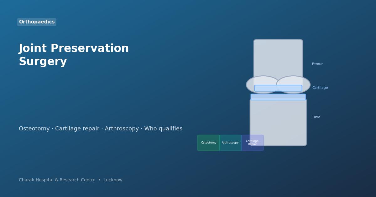
What Is Joint Preservation?
Joint preservation is a spectrum of surgical and non-surgical strategies designed to protect the joint from further damage, correct problems that accelerate wear, and restore function — all while keeping the patient's own bone and cartilage intact.
It is particularly important for younger, active patients whose knee or hip damage would typically require a joint replacement — but who are too young for an implant (which has a limited lifespan and may need revision surgery after 15–20 years).
Who Is a Candidate?
Joint preservation is most appropriate for:
Joint replacement implants last 15–20 years — preserving the natural joint avoids an early revision surgery.
Sports injuries — ACL tears, meniscus damage, cartilage lesions — can be repaired rather than replaced.
When only one side of the knee is worn (medial or lateral), realignment surgery can offload that side.
Meniscus tears, cartilage defects, ligament injuries — addressed early, these respond well to preservation surgery.
Key Preservation Procedures
Diagnostic & Therapeutic Arthroscopy
A camera (arthroscope) is inserted through small incisions to visualise the joint interior. Most joint preservation procedures are done arthroscopically — with minimal surgical trauma, faster recovery, and lower infection risk than open surgery.
High Tibial Osteotomy (HTO)
The shinbone (tibia) is cut and repositioned to shift body weight away from the damaged inner compartment of the knee to the healthier outer compartment. This reduces pain, slows further cartilage loss, and can delay knee replacement by 10–15 years in suitable patients.
Microfracture / OATS / ACI
Three techniques for cartilage repair: Microfracture makes tiny holes in bone to stimulate stem-cell-rich blood clot formation. OATS (Osteochondral Autograft Transfer) transplants a plug of healthy cartilage from a non-weight-bearing area to the damaged site. ACI (Autologous Chondrocyte Implantation) grows the patient's own cartilage cells in a lab and implants them.
ACL / PCL / LCL Reconstruction
Torn knee ligaments — especially the ACL — destabilise the joint and accelerate cartilage wear. Reconstruction using a graft (from the patient's own hamstring, patellar tendon, or a donor) restores joint stability, prevents further damage, and allows return to sports.
Meniscus Repair vs. Meniscectomy
The meniscus acts as a shock absorber in the knee. When torn, the priority is always repair (meniscus repair) rather than removal (meniscectomy) — because losing the meniscus accelerates arthritis. Modern arthroscopic techniques allow repair of tears that were previously removed.
Hip Arthroscopy & Femoroacetabular Impingement (FAI) Correction
FAI is a condition where abnormal bone shape causes the hip to pinch during movement — damaging the labrum and cartilage. Hip arthroscopy corrects the bone shape and repairs the labrum, preventing early hip arthritis and preserving the natural hip joint.
Arthroscopy — The Keyhole Advantage
Most joint preservation procedures at Charak Hospital are performed arthroscopically — through incisions just 5–10mm in size. Compared to open surgery, arthroscopy offers:
- Less blood loss and reduced infection risk
- Shorter hospital stay (often day-care or overnight)
- Faster rehabilitation and return to activity
- Smaller scars and better cosmetic outcome
- Direct visualisation of all joint structures simultaneously
Joint Preservation vs. Joint Replacement
| Factor | Joint Preservation | Joint Replacement |
|---|---|---|
| Best age group | Under 55–60 | Over 60 (or when preservation fails) |
| Implant | None — natural joint kept | Metal/plastic implant |
| Sports/high activity | Usually allowed after recovery | Restricted (implant wear) |
| Recovery time | 2–6 months (procedure-dependent) | 3–6 months |
| Longevity | 10–15+ years when successful | 15–20 years (may need revision) |
| Suitability | Partial damage, young patients | End-stage arthritis, elderly |
Recovery & Outcomes
Recovery varies by procedure. Arthroscopic meniscus repair: 6–8 weeks to return to walking normally; 4–6 months to sports. HTO osteotomy: 3–6 months for return to full activity. ACL reconstruction: 9–12 months to competitive sport. Most patients report significant pain relief and improved function within 3 months.
Consult Charak Hospital's Joint Specialist
Dr. Imran Akhtar, Orthopaedic Surgeon & Arthroscopy Specialist, offers comprehensive joint assessment and preservation surgery at Charak Hospital Lucknow. See our Orthopaedic & Joint Replacement Department for the full range of procedures available.